
Giant Odontoma with Dozens of Denticles
Giant odontomas are infrequently encountered in clinical practice. To increase familiarity with this lesion, Brooks et al present a case report with radiographic and surgical images of a compound odontoma in the mandibular anterior region.
Case Report
An otherwise healthy 21-year-old woman was referred for evaluation of a large, painless mass in the anterior mandible. The panoramic radiograph revealed a 61 × 37-mm mixed lesion with a well-circumscribed radiolucent rim and well-corticated borders (Fig 1). Other findings included displacement of proximate teeth, an overretained primary canine, an impacted permanent canine, and painless expansion of the alveolar bone. The preliminary radiographic diagnosis was compound odontoma.
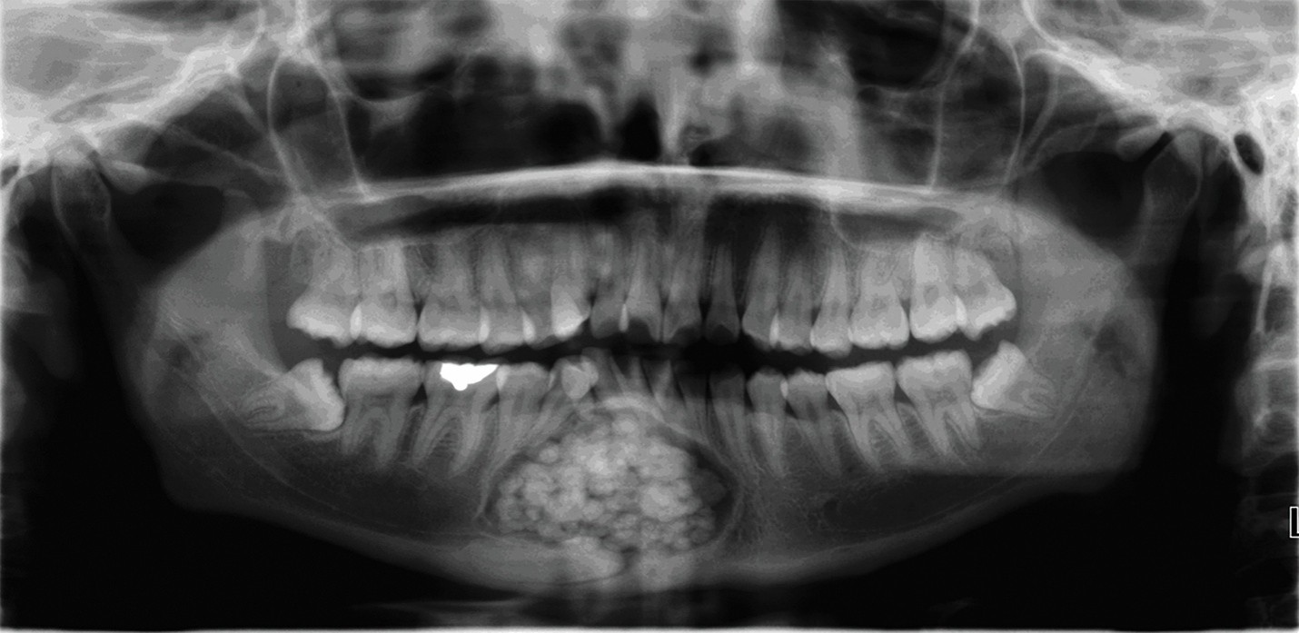
The patient was referred for surgical removal of the lesion, and the mass was easily enucleated in a single piece. The surgical specimen was 30 mm long and contained numerous tooth-like structures. Histologic findings consistent with a compound odontoma included enamel matrix with a basophilic fish scale appearance, areas of dentinoid material with abundant lamellar (Liesegang ring) calcifications, and scattered strands of odontogenic epithelium. Six months postoperatively, radiographs demonstrated osseous regeneration of the surgical site.
Discussion
Four subtypes of odontoma have been described: compound, complex, hybrid, and peripheral (oral mucosal).1-3 Larger odontomas can promote tooth malpositioning, tooth impaction, pain, bony swelling, and facial distortion. Giant odontomas are most likely to be associated with first and second molars and are less frequently adjacent to an impacted canine, as seen in this case.
Histopathologically, compound odontomas are composed of calcified tooth-like aggregations (denticles), ranging from a few to more than 500, while complex odontomas are characterized by mature dentin admixed with miniature islands of immature enamel.1 Radiographically, the compound variant demonstrates a pronounced mixed appearance due to the tooth-like structures, while the complex variant is predominately radiopaque and without resemblance to teeth.4 The radiographic differential diagnosis of odontoma should include osteoma, mature ameloblastic fibro-odontoma, and impacted tooth.
In the featured case, the findings were consistent with a giant compound odontoma, and a radiographic estimate totaled at least 52 denticles (Fig 2). Panoramic radiographs often magnify objects in the anterior region, explaining the discrepancy between the radiographic measurement (61 mm) and actual size (30 mm) of the lesion.
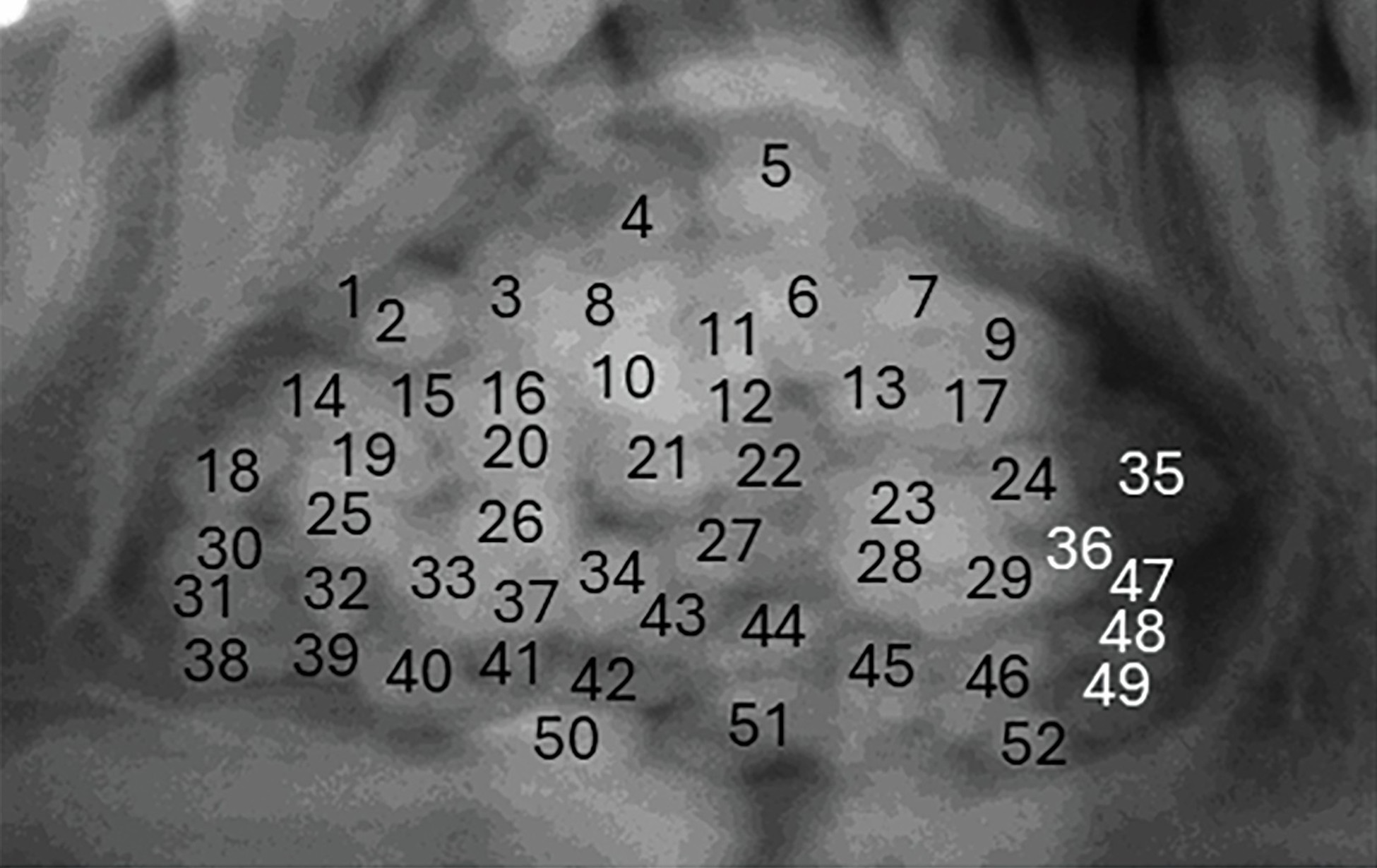 Fig 2. Compound odontoma that includes at least 52 tooth-like structures.
- Muller S, Odell EW, Tilakaratne WM. Odontogenic and maxillofacial bone tumours. In: WHO Classification of Tumours Editorial Board, ed. Head and Neck Tumours. Part A. WHO Classification of Tumours. 5th ed. Vol 9. International Agency for Research on Cancer; 2024:348-349.
- Brooks JK, McAuley PK, Qazi MJ, Sultan AS. Painful and partially erupted odontoma: a case report. Gen Dent. 2026;74(1):62-66.
- Soluk-Tekkeşin M, Balkan B, Akatay DK, Atalay B. A rare case of peripheral compound odontoma and review of the literature. Head Neck Pathol. 2022;16(3):913-917. doi:10.1007/s12105-022-01421-0
- Cheng FC, Yu-Fong Chang J, Chen MH, et al. Radiographic characteristics of odontomas in patients in the National Taiwan University Children’s Hospital. J Dent Sci. 2023;18(1):392-399. doi:10.1016/j.jds.2022.10.020
- Brooks JK, Tran LT, Basile JR, Khoury ZH, Wu LL, Price JB. Synchronous gubernacular canals with compound odontoma associated with a calcifying odontogenic cyst and transmigrated canine: an extremely rare event. Pediatr Dent J. 2020;30(1):39-44. doi:10.1016/j.pdj.2019.12.003
- Alarcón Apablaza J, Muñoz G, Arriagada C, Bucchi C, Masuko TS, Fuentes R. Odontoma recurrence. The importance of radiographic controls: case report with a 7-year follow-up. Medicina (Kaunas). 2024;60(8):1248. doi:10.3390/medicina60081248